Contact Us
Schedule your appointment online or give us a call to get started today.

Digital radiography replaces traditional film with electronic sensors and computer systems to capture dental images. Instead of waiting for film to be developed, images are available instantly on a monitor, which transforms how clinicians evaluate oral health. This modern approach streamlines diagnostics and keeps the focus on accurate, timely care rather than on analog processing steps.
Because the image is created and stored as a digital file, it can be enhanced, archived, and retrieved without degradation over time. Clinicians can adjust contrast, zoom in on areas of interest, and compare side-by-side images to track changes between visits. Those features make it easier to detect subtle issues early—often before symptoms appear—and to explain findings clearly to patients.
At Black Mountain Family Dentistry, we rely on digital radiography to support evidence-based decisions and to involve patients in their treatment planning. The technology supports a more informed conversation about oral health by providing crisp visuals that both clinicians and patients can review together.
One of the most compelling benefits of digital radiography is a meaningful reduction in radiation exposure compared with conventional film x-rays. Improvements in sensor sensitivity and image processing allow high-quality diagnostic images to be obtained with less radiation while still meeting safety standards. This reduction is particularly valuable for preventive care and routine monitoring when imaging is performed periodically.
Beyond the lower exposure, digital workflows minimize the handling of chemical developers and paper, creating a cleaner, more environmentally friendly process. Shorter imaging times also reduce the need for repeat shots, which further decreases cumulative exposure and enhances patient comfort—especially helpful for children and people who find staying still for long periods difficult.
Safety remains a priority throughout the imaging process. Digital radiography systems are used in conjunction with established protective practices and protocols, and images are reviewed immediately so any questions about image quality can be addressed on the spot.
Digital sensors capture images with fine detail that can be enhanced through software tools, improving the detection of cavities, bone changes, and other conditions that might be hard to see on older film. Adjustments for brightness, contrast, and magnification help clinicians identify issues at an earlier stage, which supports conservative treatment approaches and better long-term outcomes.
Because images display instantly, the care team can review findings together and consult in real time. This collaborative capability speeds the diagnostic process and reduces uncertainty. It also makes interdisciplinary care simpler when cases require input from specialists or collaboration with labs, since files can be shared quickly and securely.
High-quality digital images also improve record-keeping. Clear, consistent images make it easier to monitor changes over time—useful for tracking the progression of a condition or evaluating the outcome of a treatment—so both clinicians and patients have a reliable visual history of oral health.
Digital radiography is part of a connected clinical ecosystem. Because images are stored electronically, they integrate smoothly with electronic health records, treatment planning software, and other diagnostic tools. This connectivity supports streamlined workflows for procedures ranging from routine fillings to more complex restorative or implant planning.
For patient education, digital images are invaluable. When clinicians display an image on-screen and annotate the area of concern, patients gain a clearer understanding of what is happening in their mouths and why a particular treatment is recommended. That visual context often leads to better engagement and more informed decisions about care.
In practices that use multiple digital modalities, radiographs can be combined with intraoral photos and other scans to build a comprehensive portrait of a patient’s oral health. This layered approach gives clinicians multiple perspectives on an issue, which can enhance precision when designing treatment plans.
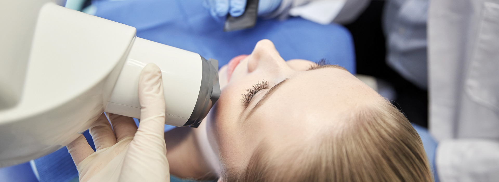
The process for a digital dental x-ray is quick and straightforward. A small sensor or plate is placed in the mouth while a handheld x-ray unit or wall-mounted device captures the image. The positioning is similar to traditional film techniques, but the workflow is optimized to take fewer images and obtain useful diagnostic information efficiently.
After the image is taken, it appears on a computer screen within seconds. The clinician will review the image with you, pointing out any areas of interest and explaining how the findings relate to your oral health. If additional views are needed, they can be taken immediately, which avoids the delays that can occur with film-based systems.
Patients generally find the experience comfortable and unobtrusive. If you have concerns about imaging, bring them up during your visit—your care team can explain the safeguards in place and how digital radiography contributes to a safer, more precise diagnostic process.
In summary, digital radiography offers faster imaging, enhanced diagnostic capability, and improved safety compared with traditional film techniques. It integrates with modern dental systems to support clear communication, accurate treatment planning, and ongoing monitoring of oral health. For more information about how digital imaging is used in our practice and what it can reveal about your dental health, please contact us to speak with a member of our team.
Schedule your appointment online or give us a call to get started today.
